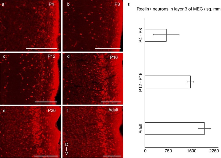
Figure 4. Increase of reelin expression in layer 3 neurons of MEC through development.
Parasaggital sections of the MEC processed for reelin-immunoreactivity (red). The sections show reelin+ stellate cells in layer 2 and an increasing reelin expression in layer 3 neurons with development in (a) P4 rat. (b) P8 rat. (c) P12 rat. (d) P16 rat. (e) P20 rat. (f) Adult rat. (g) Increasing density of reelin+ neurons in layer 3 of MEC from P4-P8 (n=1405 neurons, 4 rats); to P12-P16 (n=3309 neurons, 3 rats) to adults (n=5039 neurons, 3 rats). Error bars denote SD. Scale bars 250 µm. D- Dorsal; V- Ventral. Orientation in (f) applies to all sections.
DOI: http://dx.doi.org/10.7554/eLife.13343.010

