. 2016 Apr 2;5:e13343. doi: 10.7554/eLife.13343

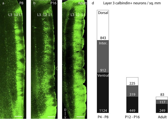
Figure 5. Dorsal-to-ventral disappearance of layer 3 calbindin expression.

Parasaggital sections showing superficial layers of the MEC processed for calbindin-immunoreactivity (green). (a) Calbindin expression is seen throughout layer 3 in P8 rats. (b) Calbindin expression is seen only in ventral half of layer 3 in P16 rats. (c) Calbindin expression is largely absent in layer 3 in adult rats. (d) Proportion of layer 3 calbindin+ neurons in dorsal (white), intermediate (gray) and ventral (black) MEC in P4-P8 (n=3776 neurons, 8 rats); P12-P16 (n =2014 neurons, 8 rats); and adult (n=828 neurons, 7 rats) rats. The numbers represent layer 3 calbindin+ neuronal density and decay in a dorsal to ventral gradient with age as evident with the reduced proportions of the white (dorsal MEC) and gray (intermediate MEC) sections of the columns with increasing age. Scale bars 250 µm. L1- Layer 1; L2- Layer 2; L3- Layer 3; D- Dorsal; V-Ventral. Orientation in (c) applies to all sections.

DOI: http://dx.doi.org/10.7554/eLife.13343.012

Figure 5.

Figure 5—figure supplement 1. Dorsal- ventral distribution of layer 3 reelin expression.

Figure 5—figure supplement 1.

Parasaggital sections showing superficial layers of the MEC processed for reelin-immunoreactivity (red). (a) Reelin expression is sporadic throughout layer 3 in P8 rats. (b) Reelin expression equitably increases in layer 3 in P16 rats. (c) Reelin expression is present throughout layer 3 in adult rats. (d) Proportion of layer 3 reelin+ neurons in dorsal (white), intermediate (gray) and ventral (black) MEC in P4-P8 (n=1405 neurons, 4 rats); P12-P16 (n =3309 neurons, 3 rats); and adult (n=5039 neurons, 3 rats) rats. The numbers represent layer 3 reelin+ neuronal density and increase equitably with age as evident with the similar proportions of the white (dorsal MEC), gray (intermediate MEC) and black (ventral MEC) sections of the columns with increasing age. Scale bars 250 µm. L1- Layer 1; L2- Layer 2; L3- Layer 3; D- Dorsal; V-Ventral. Orientation in (c) applies to all sections.
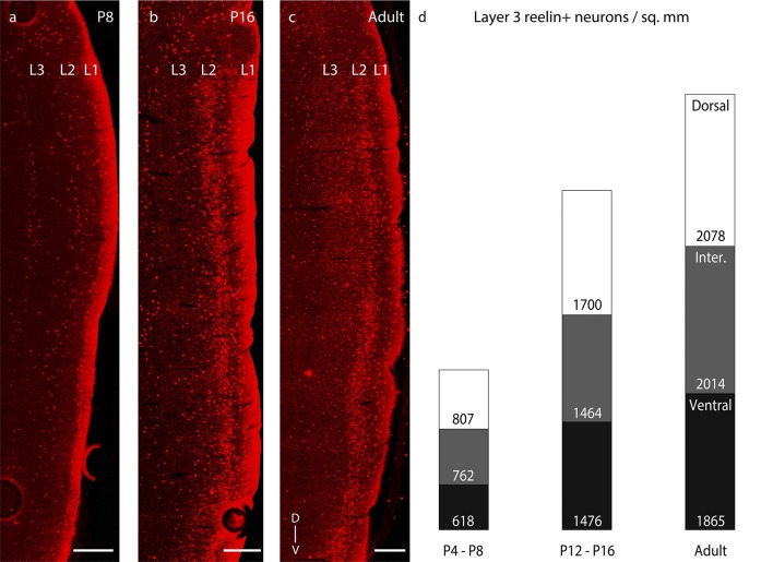
Figure 5—figure supplement 1—source data 1. Calbindin+ neurons (Figure 5) and reelin+ neurons (Figure 5—figure supplement 1) counted and areas (in μm2) in dorsal, intermediate and ventral parts of layer 3 for determining calbindin+ and reelin+ neuronal densities respectively in P4-P8, P12-P16 and adult rats.
DOI: 10.7554/eLife.13343.014