. Author manuscript; available in PMC: 2016 Aug 1.
Published in final edited form as: J Magn Reson Imaging. 2014 Dec 4;42(2):269–279. doi: 10.1002/jmri.24802

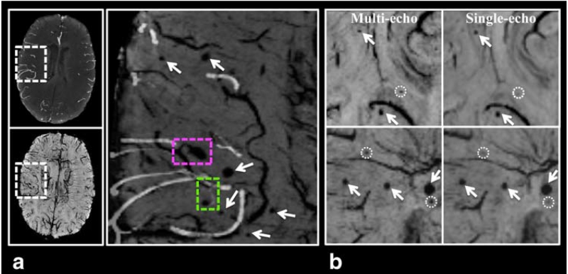
FIGURE 6. Benefits from using the multi-echo sequence. A: The multi-echo sequence allows arteries from TOF-MRA images (maxIP over 8 mm) to be overlaid on CMBs and veins from minIP SWI images without the need for registration. From the overlaid image (zoomed in to a local region specified by the dashed-box on the original images) on the left column, it can be clearly seen that one CMB is arising from venous vessels (pink dashed-box) while another one appears to originate from arterioles (green dashed-box). B: minIP SWI Images from two different patients (top and bottom rows) showing CMBs (arrows) that are visualized on both single- and multi-echo images, and three additional CMBs that are only detected on multi-echo SWI (dashed-circles).

FIGURE 6