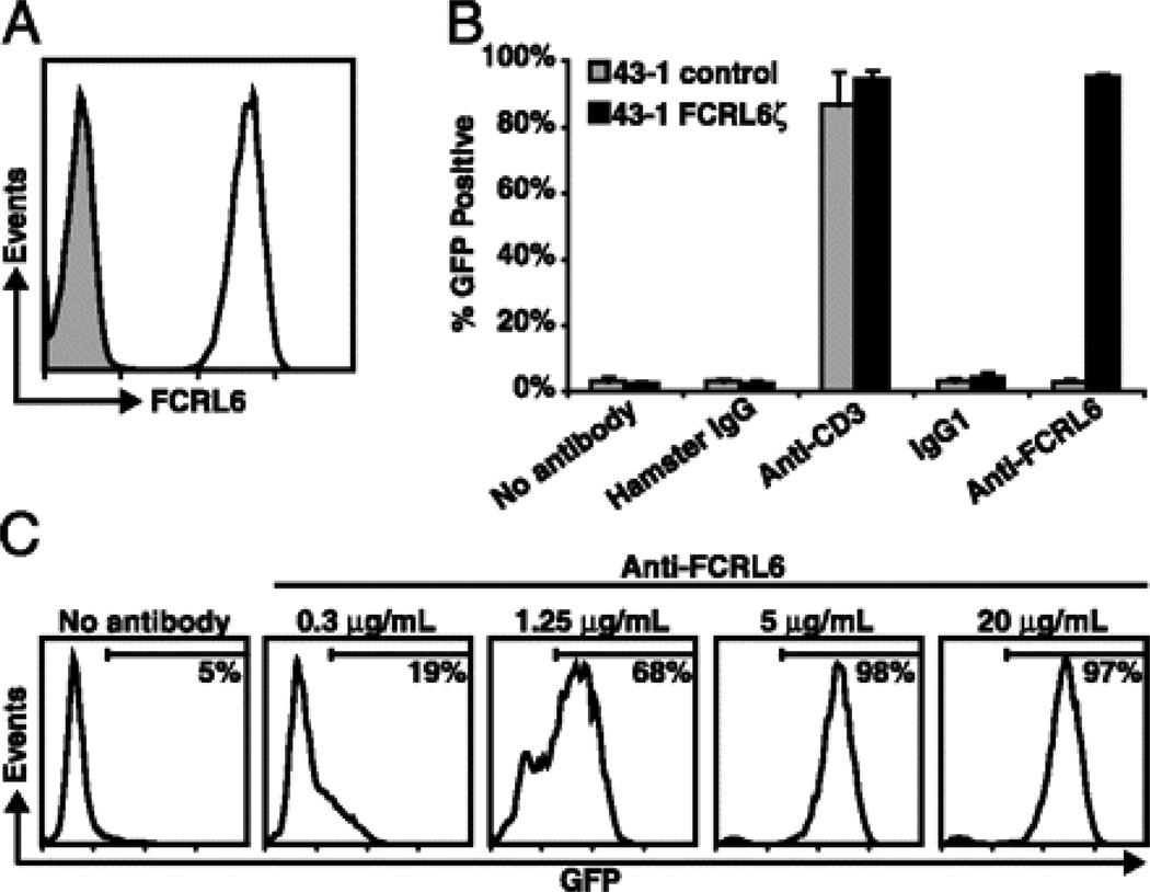
FIGURE 1.
Development of a GFP-inducible system to detect FCRL6 engagement. A 43-1 cells transduced with the FCRL6ζ reporter construct were stained with the anti-FCRL6 7B7 mAb (black line) or with an isotype-matched control (grey shade). B Untransduced 43-1 control (grey columns) or 43-1 FCRL6ζ cells (black columns) were stimulated at 5µg/mL with the indicated plate-bound antibodies for 18 h and GFP induction was assayed by flow cytometry. Columns represent the mean ± s.d.; n=3. C FCRL6ζ cells were stimulated as in panel (B) with different concentrations of plate-bound anti-FCRL6 mAb. The percentage of GFP+ 43-1 cells is indicated in each histogram and demarcated by horizontal bars.

