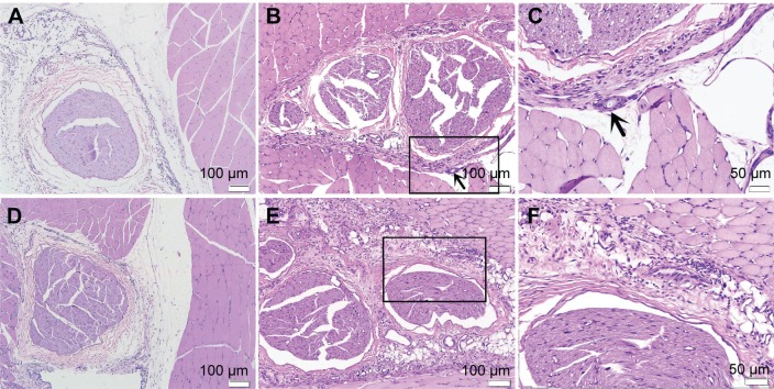
Figure 4.
Sciatic nerves and surrounding tissues were stained with hematoxylin and eosin at 7 d after administration.
Notes: (A) Image of rats in control group, no inflammation cell was observed (×100). (B) Image of rats treated with PELA, some inflammation cells were observed (×100). (C) High-magnification images of (B), arrows to the inflammation cells and microparticles (×200). (D) Image of rats in Rop group, slight inflammation was observed near the sciatic nerve (×100). (E) Image of rats in Rop-PELA group, granulation tissue and moderate degree inflammation were detected at the site of administration (×100). (F) High-magnification images of (E) (×200). Sciatic nerves in all groups were almost normal.
Abbreviations: Rop, ropivacaine; PELA, poly ethylene glycol-co-poly lactic acid; d, day.

