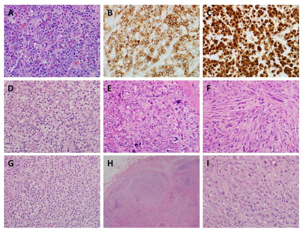
Figure 1. Pathological features of ALK-positive ALCL.
(A) Photomicrograph of typical ALK-positive ALCL, common pattern, showing sheets of large atypical lymphocytes (“hallmark” cells). (B) Immunohistochemical staining for CD30 in the same case shows the tumor cells to be positive, with a membranous and Golgi pattern of staining. (C) The tumor cells are positive for ALK. The presence of both nuclear and cytoplasmic staining indicates the ALK fusion partner is likely to be NPM. (D) Lymphohistiocytic pattern, showing tumor cells interspersed in a background of small lymphocytes and histiocytes. (E) This example shows a predominance of very large, markedly pleomorphic cells. (F) Case with so-called “sarcomatoid” features, showing marked spindling of the tumor cells. (G) Small cell variant, showing mostly small tumor cells with abundant pale cytoplasm. (H) Low-power image of case with “Hodgkin-like” features, including tumor nodules separated by bands of sclerosis. (I) Despite the Hodgkin-like appearance at low power, a higher power image shows sheets of hallmark cells typical of ALCL.

