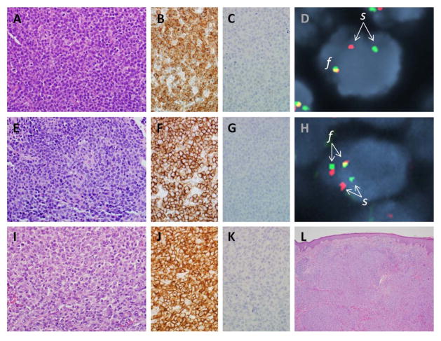
Figure 2. Pathological features of ALK-negative ALCL.
(A) Photomicrograph of ALK-negative ALCL with DUSP22 rearrangement (see text), showing sheets of tumor cells. (B) Tumor cells from the same case are positive for CD30 by immunohistochemistry. (C) Tumor cells are negative for ALK. (D) Fluorescence in situ hybridization (FISH) using a breakapart probe to the DUSP22-IRF4 locus on 6p25.3 shows a tumor cell nucleus (blue counterstain) with a single normal fusion signal (f) on one allele and abnormal separation (s) of the red and green portions of the probe on the other allele, indicating the presence of a rearrangement. (E) ALK-negative ALCL with TP63 rearrangement (see text). (F) Tumor cells are positive for CD30. (G) Tumor cells are negative for ALK. (H) FISH using a dual-fusion probe to the TBL1XR1 and TP63 loci on 3q show normal separation (s) of the two loci on one allele, and TBL1XR1-TP63 fusion (f) on the other allele. (I) Primary cutaneous ALCL, showing histological features similar to those of both ALK-positive and ALK-negative systemic ALCL. (J) Tumor cells also show a similar pattern of staining for CD30. (K) Tumor cells are negative for ALK. (L) A low-power image shows the relationship of the tumor cell infiltrate to the overlying epidermis.

