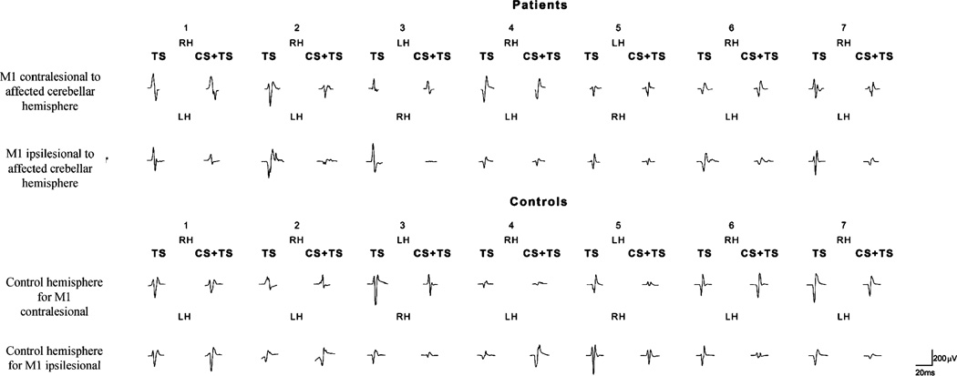
Fig. 2.
Raw data, motor-evoked potentials after administration of test stimulus (TS) or paired pulses (conditioning stimulus, CS, followed by test stimuli, TS) in the right (RH) and left (LH) hemispheres in patients and controls. In patients, decreased inhibition of test responses is noticed in cerebral hemispheres contralateral to cerebellar infarcts compared to hemispheres ipsilateral to cerebellar infarcts. In healthy controls, inhibition of test responses were similar in the two cerebral hemispheres

