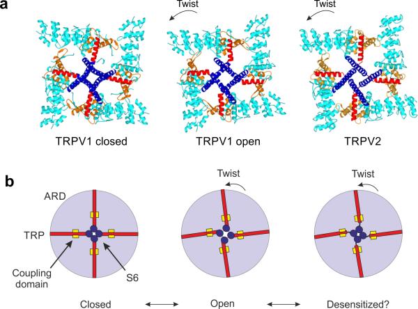
Figure 6.
Coupling between the TRP domain and S6. (a) Top view of the closed TRPV1, open TRPV1 and TRPV2 structures. S1–S5 have been removed for simplicity. Rotation of ARD (cyan) leads to a displacement of the TRP domain (red), which pulls on the S6 (blue) and thereby drives the opening of the lower gate. In TRPV2, the coupling between S6 and the TRP domain is disrupted, and the ARD rotation is therefore not translated to an opening of the channel. (b) Cartoon of the proposed gating mechanism in TRPV channels. The rotation of ARD (light blue circle) is directly coupled to opening of the lower gate through the TRP domain (red). This coupling is guided by the pre-S1 helix, the linker domain and CTD, which we collectively refer to as the ‘coupling domain’ (yellow square).

