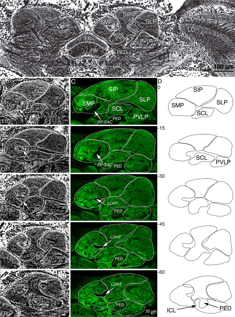
Figure 2.
Divisions of the superior protocerebrum. A: Part of a frontal section stained by the Bodian method showing the location of the fan-shaped body (FBu, FBl) and volumes of the superior protocerebrum. Shown are the superior median, superior intermediate, and superior lateral protocerebrum (SMP, SIP, SLP). These lie above the mushroom body pedunculus (PED) here seen in cross-section. The underside of the PED is flanked by the inferior clamp neuropil (ICL, lower image, panel D). The fan-shaped body lies beneath the pars intercerebralis, and central to the paired lateral accessory lobes (LAL). The optic lobe’s lobula (LO) is shown to the right. Axons from the optic lobe reach the central brain via the optic pedunculus (OP PED). B: Five panels showing a sequence of 15-µm-thick frontal sections (0 µm, the most frontal of the series to −60 µm, the most posterior) through the superior protocerebrum to illustrate gradual changes of protocerebral volumes through a depth of 60 µm. The anatomical divisions of the superior protocerebrum are outlined and are shown separately in panel D. C: A corresponding series of vibratome sections labeled with an antiserum raised against synapsin. In both B,C, the white arrows indicate a tract of axons occupied by neurons connecting the superior protocerebrum with the fan-shaped body (superior protocerebrum–superior arch commissure SP-SAC: axons shown in Figs. 3–7). The laterodorsal horizontal tract (LDHT) is shown at level −30 to −60 in panel C. D: Outlines of divisions of the superior protocerebrum, obtained by averaging traces from B,C, showing the arrangements among the discussed neuropil regions. [Color figure can be viewed in the online issue, which is available at wileyonlinelibrary.com.]

