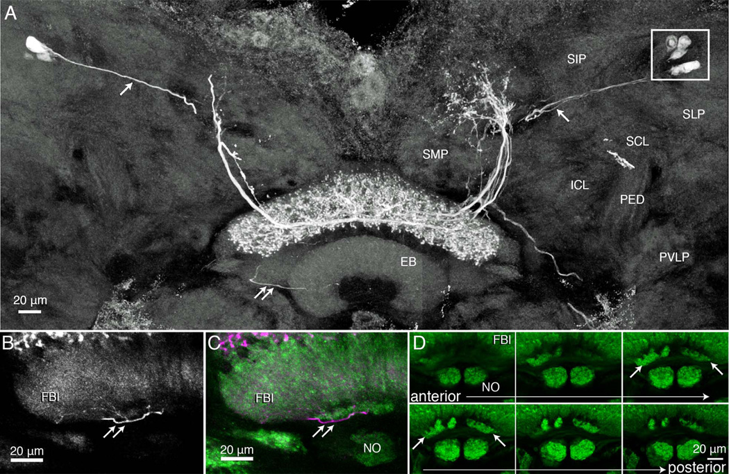
Figure 4.
Tangential neurons with terminal arborizations through all the strata (8–6) of the upper division of the fan-shaped body. A: Composite image, reconstructed from confocal stacks through two consecutive sections at the level of the fan-shaped body and the ellipsoid body (EB). Several neurons have been filled with neurobiotin after an intracellular recording and injection from one neuron. Together, these dye-coupled cells have extensive dendritic branches in an upper middle domain of the superior medial protocerebrum (see schematic, Fig. 9). Axons project from the protocerebral processes to stratum 6 of the upper fan-shaped body where they give rise to varicose and beaded processes extending upward out to, and including, stratum 8. Thin fibers (single white arrows) connect the protocerebral arborizations to cell bodies (boxed, upper right) located dorsal to the superior lateral protocerebrum (SLP). Double arrows indicate one of several thin processes that extend from the terminal arborization downward, behind the ellipsoid body and into one of the two asymmetric bodies. The terminal of one of these collaterals is shown in the enlargement of panel B. C,D: Six consecutive sections through the lower margin of the FBu, showing the noduli, and above them the asymmetric bodies within stratum 1. Other regions shown in A are the superior intermediate protocerebrum (SIP), the superior and inferior clamp (SCL, ICL), both of which enclose the mushroom body’s pedunculus (PED), and part of the posterior ventrolateral protocerebrum (PVLP).

