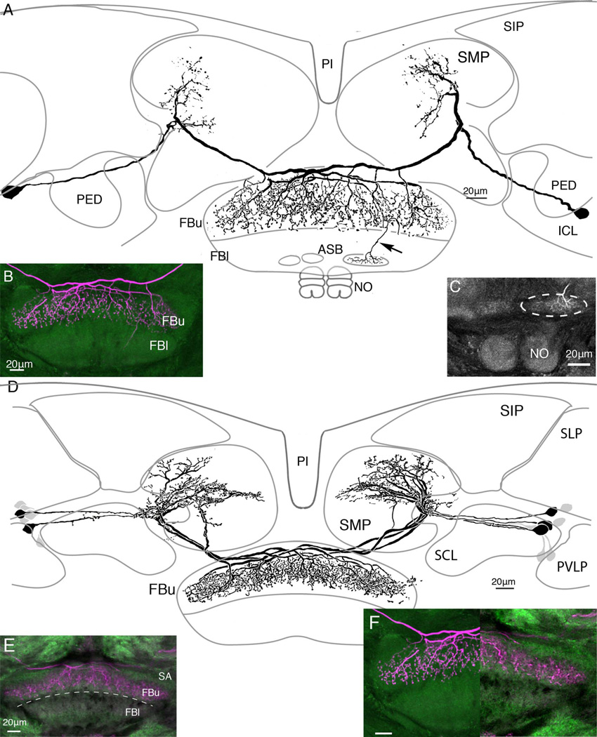
Figure 5.
There are subtle yet clear distinctions of tangential neuron morphologies. A: Paired tangential neurons with their main terminal branches above the superior arch extend long beaded collaterals through strata 8, 7, and 6. B: Confocal stack showing terminal branches that appear to hang down through the upper fan-shaped body (FBu). The distal arborizations of these neurons occupy a vertically arranged domain in the lateral part of the superior medial protocerebrum. One of the two terminals also supplies a collateral to the larger of the two asymmetric bodies (inset C, middle right). D: Reconstruction of neurobiotin-filled neurons, the terminal branches of which extend within and just beneath the superior arch (see also confocal stack, inset E). The branches provide a dense system of fine varicose processes in strata 7 and 6. F: A side-by-side comparison of the neurons shown in A,D (lower right inset, F) demonstrates their difference with respect to their depth of termination in the FBu. Neurons shown in D have their distal arborizations occupying a dorsolateral domain of the superior medial protocerebrum. After filling a single neuron with neurobiotin, a total 15 cell bodies were visible, some more intensely labeled than others.

