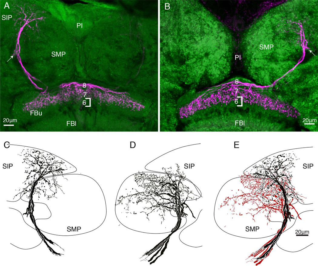
Figure 6.
A,B: As in Figure 5, here two morphological types of neurons terminate at overlapping but different depths of the upper fanshaped body. The neuron in A occupies a thin domain in stratum 7, whereas the neuron in B has processes reaching into stratum 6. Such differences are subtle, in contrast to their obvious distinctions, which refer to their branches in the superior medial protocerebrum. C,D: Neurons terminating as in panel A have most of their branches in the superior intermediate protocerebrum (SIP). Neurons terminating as in panel B have their branches in the superior medial protocerebrum (SMP). D: These distinctive domains are further accentuated when the two types of arborizations are superimposed.

