Fig. 10.
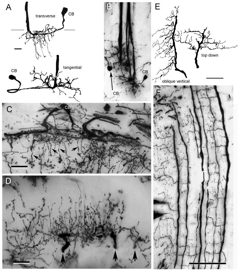
Type 2 tangential neurons. A. C. granulatus: the wide field dendrites of this cell type reside superficially in the medulla, some sending long slender branches to deeper levels (upper drawing, transverse view). Top-down views (lower drawing and panel B) show these dendrites extending radially from the axon. Neurites lead from these dendritic fields to elongated cell bodies (CB) near the medulla surface. C. H. oregonensis: transverse section of the medulla showing similar dendritic components from which arise slender processes into medullary columns (arrows). D. H. oregonensis: traverse section of the lamina showing three adjacent terminal branches of one type 2 tangential (at arrows) giving rise to a dense arrangement of varicose processes that ascend through the lamina’s plexiform layer. E. C. granulatus camera lucida drawings showing terminal arbors of two type 2 tangential neurons. F. C. granulatus: vertical section showing en masse impregnated type 2 tangential endings. Adjacent “giant” processes provide orthogonally directed collaterals from which arise ascending processes. Scale bars for A, B=10 μm, C, D = 25 μm, for E, F = 50 μm.

