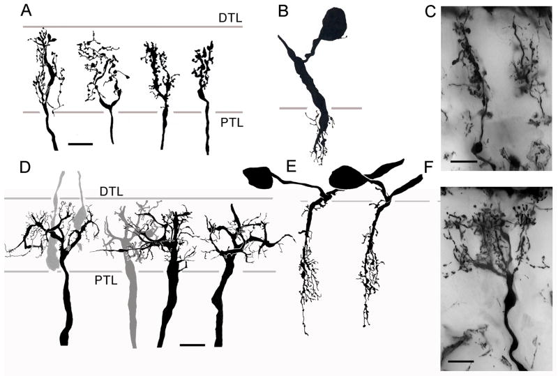
Fig. 11.
Small (Type 1) and wide-field (Type 2) T-cells. Four variants of type 1 T-cell dendritic arbors in C. granulatus. Processes originate from two, almost parallel processes in the lamina that arise from a slender axis fiber. These give off many small varicose and pinhead-like specializations. B. The assumed medulla component of these T-cells consist of a swelling from the axon that tapers to provide several short branches within the outer strata of the neuropil. A very short neurite links this component to the soma, which is located just above the medulla. C. Homologous dendritic trees in the lamina of H. oregonensis. D. Four different views of Type 2 T-cell dendrites in C. granulatus. The axon gives rise to several stout V-shaped divisions that provide a system of short spine-like specializations. The dendritic trees extend through several optic cartridges. E. Their medulla components consist of narrow cascades of spiny processes connected by a neurite to somata located just above the medulla. F. Type 2 T-cell dendrites in H. oregonensis give rise to clusters of specializations, each cluster located at an optic cartridge. All scale bars = 10 μm.

