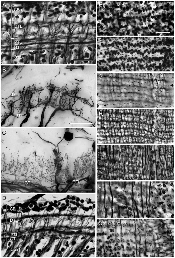
Fig. 2.
Neuroarchitecture of the lamina as revealed by Bodian’s reduced silver with Golgi impregnations to show selective neuron correlates. A. H. oregonensis: Section cut parallel to the vertical axis of the retinotopic mosaic showing levels depicted top-down in panels E–K. An outer cell body layer (CBo, and panel E) provides monopolar cells with long neurites such as the M2 type shown in panel C. The inner layer of cell bodies (CBi, and panel F) provides monopolar cell M1 and M5. A short intermediate zone reveals the clustered neurites of monopolar cells (panel G). The distal tangential layer (DTL, also panel H) receives contributions from the bilayered terminals of the type 1 tangentials (panel B) with the same cell type providing tangential processes of the proximal tangential layer (PTL and panel I). The “giant” tangential layer (GTL) is provided by the primary branches of type 2 tangential endings (panel C and J). The bundled axons from optic cartridges that provide the first optic chiasma (Och1, also panel K) originate just beneath this layer. D. Bodian stained section of the lamina of C. granulatus shows the same layering except for the monopolar cell perikarya, where the two layers are virtually indistinguishable. Scale bars for panels A–C and E–K = 25 μm; for panel D = 50 μm.

