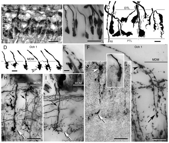
Fig. 4.
Photoreceptor terminals. A. H. oregonensis: Texas red-dextran fills show receptor terminals arranged as two tiers. B. H. oregonensis: horizontal section showing Golgi-impregnated terminals “end-on”. Note that some terminals occupy intermediate positions with respect to the tiers shown in panel A. C. C. granulatus: camera lucida drawings of receptor endings aligned along the vertical axis of the retinotopic mosaic. Spatulate endings (left and center in panel) correspond approximately to the two tiers shown from H. oregonensiswhereas other terminals (leftmost, four to right in panel, and grey terminal center) occupy intermediate positions or extend through the whole depth of the plexiform layer. The axon of a thoroughgoing R8 photoreceptor is shown extreme left. D, E. C. granulatus: swellings of R8 photoreceptors at the medulla surface. F. H. oregonensis: two R8 photoreceptor endings showing their outer swelling (arrowhead) and deep extension ending as a terminal varicosity (at arrow). Inset compares the ending of an M3 monopolar cell. G. H. oregonensis: Texas red-dextran fill of a R8 photoreceptor endings showing the surface swelling (arrowhead) and swollen terminal (at arrow). H, I. H. oregonensis: medulla terminals of M2 and M2d monopolar cells. DTL: distal tangential layer, PTL: proximal tangential layer, Och 1: level of the first optic chiasma lamina, MDM: medulla’s distal margin, OT: outer terminals, IT: inner terminals. Scale bar for panels A, B = 25 μm; panel C, =25 μm; panel D, E = 10 μm; panels F–I = 25 μm.

