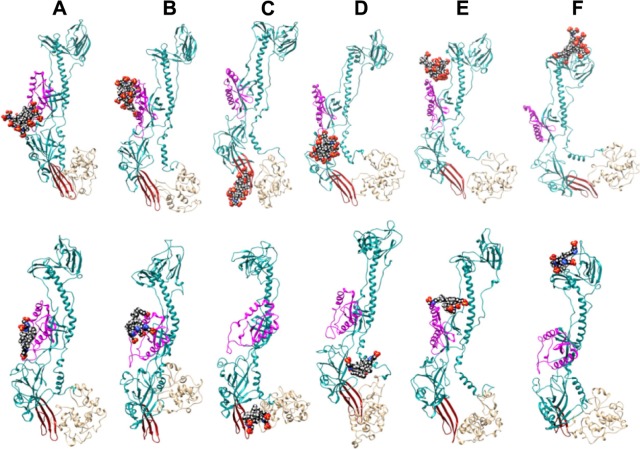
Figure 4.
Final G2-S16/gB (top) and G1-S4/gB (bottom) complexes obtained from the initial configurations (A–F) – see Supplementary materials.
Notes: The main part of the supposed binding interface for the gH–gL is highlighted in magenta. The putative fusion loops are in red and the C-terminal part comprising residues 722–904 is in tan. This terminal part of the gB was not included in our template structure (4BOM), Therefore, we simulated it from the primary amino acid sequence just to “naturally” close this incomplete part. In reality, most of this gB part anchored in viral membrane has different tertiary structure. Atoms of dendrimers are colored as follows: C, black; O, red; Si, gray; S, yellow; N, blue; H, white.

