Fig. 1.
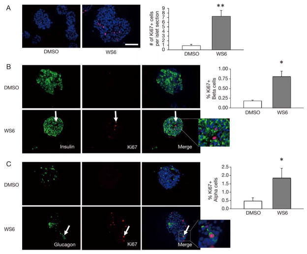
WS6 stimulates proliferation of both human pancreatic beta and alpha cells. Immunofluorescent analysis of human islets exposed to WS6 1.0 μM or DMSO (control) for 96 hours.
A. Ki67 (red) staining of human islets with quantification of total Ki67 cells per islet. B. Insulin (green) and Ki67 (red) staining of human islets with quantification of Ki67+ beta cells. White arrow shows Insulin+/Ki67+ double-positive cell.
C. Glucagon (green) and Ki67 (red) staining of human islets with quantification of Ki67+ alpha cells. White arrow shows Glucagon+/Ki67+ double-positive cell. (DAPI staining in blue. n = 4 from four different donors. Bars indicate mean ± SEM. Scale bar = 50 μm. *p < 0.05, **p < 0.01)

