Fig. 2.
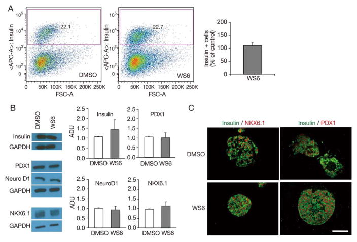
WS6 does not impact human beta cell number or beta cell differentiation. Analysis of human islets exposed to WS6 1.0 μM or DMSO (control) for 96 hours.
A. Flow cytometry analysis. Gating strategy for insulin positive cells (left) and quantification of the proportion of insulin positive cells (right) (n = 3 from three different donors, p = NS). B. Western blot analysis of human insulin and beta cell transcription factors, with representative images (left) and quantification (right, n = 3 to 4 from four different donors, p = NS for all). C. Immunofluorescent analysis showing similar NKX6.1 and PDX1 expression in DMSO and WS6 treated islets (ADU = Arbitrary density units. Bars indicate mean ± SEM. Scale bar = 50 μm). Representative images from Figs. 2A and 1B come from different donors.

