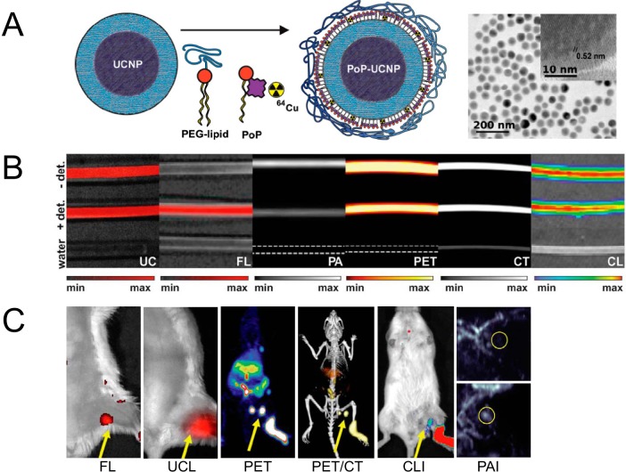
Figure 5.
Hexamodal imaging with radioactive nanomaterials. Schematic structure and transmission electronic microscopy (TEM) images of this porphyrin-/lipid-wrapped upconversion nanoparticles (UCNPs) (A). Imaging studies with material-filling tubing by upconversion luminescence (UCL), fluorescence, PAI, PET, computed tomography (CT), and Cerenkov luminescence imaging (CLI) (B). Signal intensity–tissue depth relationship was also examined. (Note: +/− det means cover or remove turkey breast over the tubing). In vivo LN mapping by these 6 imaging modalities (C). Photoacoustic (PA) images before and after the material injection are shown. Reproduced with permission from Rieffel et al (103).

