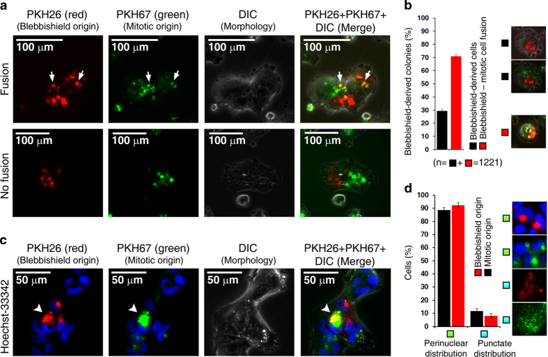
Figure 1.
Fusogenic membranes from the perinuclear region mediate fusion of blebbishields with mitotic cells. (a) Surface membranes of blebbishields and mitotic cells labeled with PKH dyes accumulated at the perinuclear region after reattachment to the substratum. Arrows indicate fusion of blebbishields with mitotic cells. (b) Quantification of blebbishield–mitotic cell fusion. n, number of colonies. (c) Blebbishield surface membranes were restricted to the trans perinuclear region, whereas mitotic-cell surface membranes occupied both the cis and trans perinuclear regions (arrowheads). DIC, differential interference contrast. (d) Blebbishield and mitotic-cell surface membranes exhibited fragmentation and perinuclear reassembly phenotypes similar to those of Golgi membranes.

