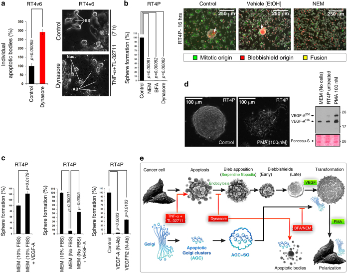
Figure 4.
Endocytosis, VEGF signaling, and Golgi integrity drive blebbishield formation and transformation from blebbishields. (a) Blocking endocytosis by dynasore promoted formation of individual apoptotic bodies and prevented formation of blebbishields (also see Supplementary Movie S2). BS, blebbishields; AB, apoptotic bodies. (b) Inhibitors of endocytosis and Golgi integrity blocked transformation from blebbishields (left panel). NEM, N-ethylmaleimide; BFA, brefeldin-A. NEM blocked reattachment of both blebbishields (PKH26 (red)) and mitotic cells (PKH67 (green)) to substratum (right panel). (c) Isolated blebbishields formed more spheres with exogenous VEGF (left panel). Serum withdrawal reduced transformation from blebbishields, and exogenous VEGF rescued transformation (middle panel). Addition of 1 μg/ml neutralizing antibodies (N-Ab) to VEGF or VEGFR2 blocked transformation from blebbishields in 10% MEM (right panel). (d) Phorbol-myristic acetate (PMA) accelerated polarization of cells from blebbishield-transformed spheres (also see Supplementary Movie S4; photomicrograph). PMA enhanced VEGF secretion detected by Western blotting of conditioned media (right panel). Note that VEGF is present in FBS of MEM (lane-1). (e) Schematic showing how endocytosis drives blebbishield formation. The status of Golgi fragmentation is depicted in blue. AGC, apoptotic-Golgi clusters; SG, surface Golgi membranes. Note: Though Golgi membranes are at the surface of blebbishields (based on PKH26 staining), transmission electron microscopy indicates that internally, Golgi remains as AGC.1

