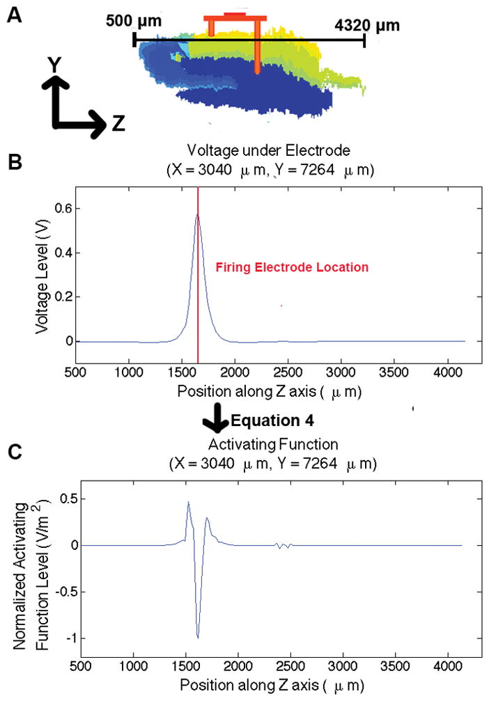
Fig. 4.

Activating function across axon located underneath the micro-electrode array. A) Slice of hippocampus model showing the location of the axon: just beneath the electrode array in the mediolateral plane, B) the voltage across the line in A, along typical axon morphology in the hippocampus, and C) the activating function applied to the voltage, showing stimulation beneath the electrode.
