
Figure 4.
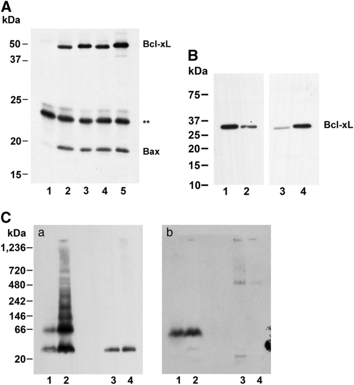
(A) Bax and Bcl-xL interaction. Binding of wild-type and S73D Bcl-xL to immobilized Bax. Bax was immobilized to anti-Bax antibody bound to Protein-G-Dynabeads and incubated with either wild-type (lanes 2, 4) or S73D (lanes 1, 3, 5) Bcl-xL. One incubation was performed without anti-Bax antibody (lane 1). Bax was obtained either from untreated cells (lanes 1–3) or from cells treated with tBid-expression adenovirus (lanes 4, 5). Bound proteins were eluted, electrophoresed by PAGE and probed for Bcl-xL. After localization of Bcl-xL, the membranes were probed for Bax. Asterisk, non-specific band from Protein-G-Dynabeads. (B) Bcl-xL on mitochondria and in cytoplasm. Bcl-xL immunoblot showing relative distribution of Bcl-xL in mitochondrial and cytoplasm fractions. Legend is the same as Figure 2c. For the cytoplasmic fraction, 10 μg protein was loaded, and for the mitochondrial fraction 1 μg protein was loaded. (C) Bcl-xL and Bax on mitochondria. Mitochondria were isolated from TKPTS cells and proteins extracted with 1% n-dodecyl-β-D-maltoside. Proteins from mitochondria (lanes 1, 2) and cytoplasm (lanes 3, 4) were electrophoresed on BN-PAGE. Cells were transduced with either wild-type (lanes 1, 3) or S73D (lanes 2, 4) Bcl-xL adenovirus and incubated 2 days after transduction before harvesting. Western blots were probed for Bcl-xL (a) or Bax (b).