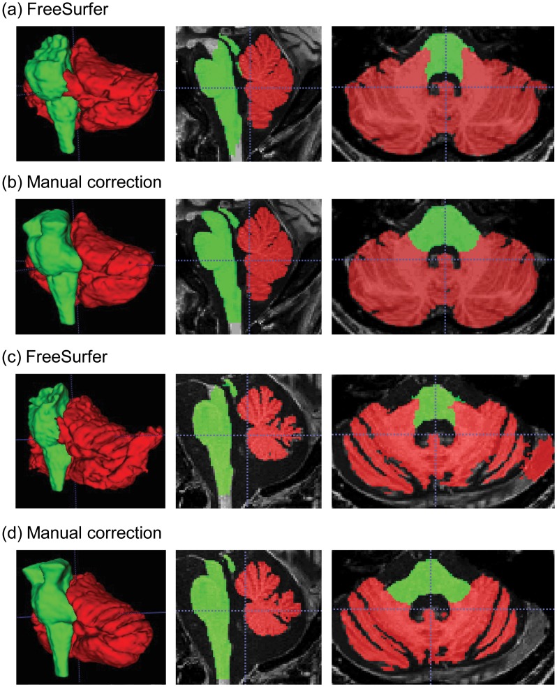
Fig 1. A sample of segmented cerebellum and brainstem.
The left column shows the 3D-reconstructed structures while the middle column show a sagittal view and the right column shows an axial view. (a) The original segmentation from FreeSurfer automated process for a healthy control; (b) the corresponding manually corrected segmentation; (c) the FreeSurfer segmentation for a patient with neurodegeneration; and (d) the corresponding manually correction segmentation. Note the top of the brainstem is filled in after manual correction as well as the correction of the cerebellar-brainstem interface and removal of non-brain tissue from cerebellar labelling. Red, cerebellum; lime, brainstem.

