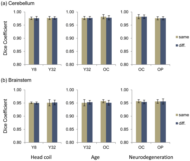
Fig 4. Dice coefficient for testing the effect of head coil used during scan acquisition and brain atrophy due to aging or neurodegenerative process on segmentation correction on (a) cerebellar segmentation and (b) brainstem segmentation.
The columns of paired bar graphs, from left to right, show the effect on Dice coefficient due head coil differences, aging, and neurodegenerative process, respectively. Y8 refers to the younger healthy control group scanned using an 8 channel head coil. Y32 refers to the younger healthy control group scanned using a 32 channel head coil. OC refers to the older healthy control group scanned using a 32 channel head coil. OP refers to the older patient group with neurodegeneration scanned using a 32 channel head coil. Error bars indicate ±1 standard deviation. “Same” indicates that the training set and the testing set contained scans from the same group; and “diff.” indicated that the training set contained scans from a comparison group of the testing set.

