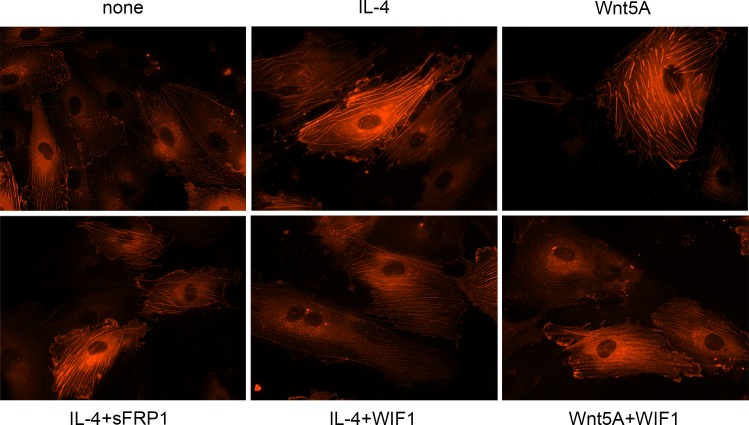
Fig 5. Stress fiber formation in IL-4 treated HCAEC.
Live actin-RFP staining showing the formation of actin stress fibers in HCAEC treated with IL-4 or Wnt5A in the absence or presence of sFRP1 and WIF1. Expression of de novo synthesized RFP-actin after transfection as outlined in methods was visually observed up to 24 h. Photomicrographs of stress fiber formation were taken randomly at 12 h after stimulation with IL-4 and Wnt5A alone or in the presence of sFRP1 and WIF1 using Zeiss Axio Observer.Z1 equipped with AxioCam MRm digital camera and ZEN 2012 software. Original magnification, 400×. Independent identical experiments in triplicates were repeated at least three times, with analogous results.

