
Figure 2.
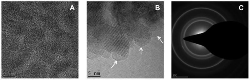
Nanocharacterization of CdTe QDs. High resolution transmission electron microscopy of CdTe QDs before and after silanization. (A) Monodisperse CdTe QDs with 5 nm mean diameter are visualized before silanization. (B) After silanization, the silica shells are identified by the low contrast amorphous outer layers (pointed by the arrows). (C) Selected area diffraction of silica-coated CdTe QDs confirms the formation of nanocrystalline materials.