
Figure 4.
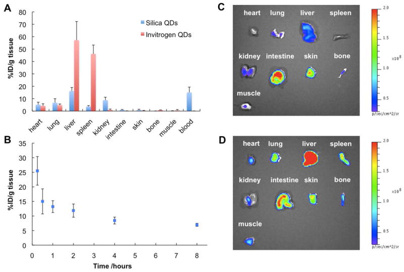
Biodistribution study of silica-coated QDs (λmax=610 nm) and Invitrogen QD605 (carboxylate). (A) Quantitative analysis of biodistribution of silica-coated QDs and Invitrogen QDs using ICP-AES. The nude mice were sacrificed 30 min after tail vein injection of QDs. QDs were completely digested using concentrated nitric acid before ICP-AES measurements. Silica-coated QDs exhibit less liver and spleen uptakes but higher kidney uptake and blood retention compared to Invitrogen QDs. Five mice were examined for each type QDs. (B) Blood retention kinetics of silica-coated QDs. Three mice were examined at each time point. (C) Fluorescence image of organs and tissues excised from the mouse injected with silica-coated QDs (shown in Figure 5A). The high signals in intestine and skin are due to autofluorescence. (D) Fluorescence image of organs and tissues excised from the mouse injected with Invitrogen QDs (shown in Figure 5B). It should be noticed that the fluorescence images are not quantitative considering the depth of tissues where QDs localized and different quantum yields of silica-coated and Invitrogen QDs.