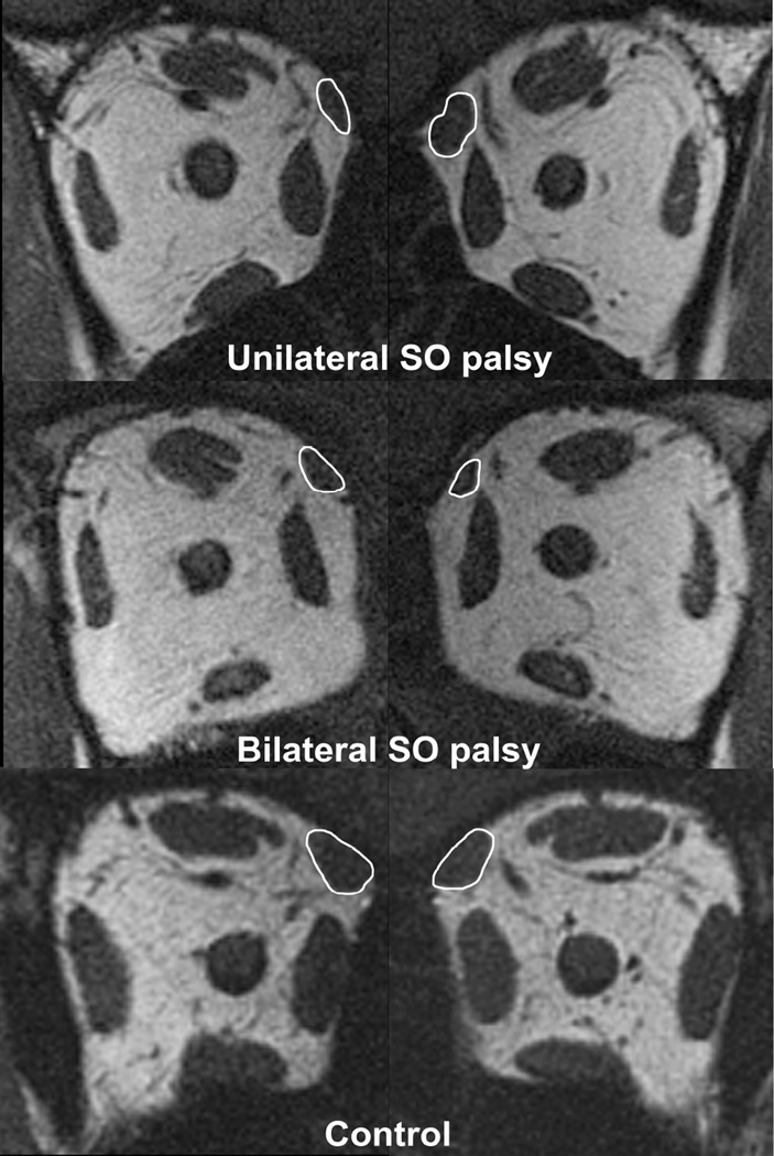
Fig. 1.
Quasicoronal MRI of right (left column) and left (right column) orbits in patients with superior oblique (SO) palsy and a normal control. Note the smaller size of the right SO muscle compared with left SO muscle in unilateral SO palsy (top row). Both SO muscles are small in bilateral SO palsy (middle row).

