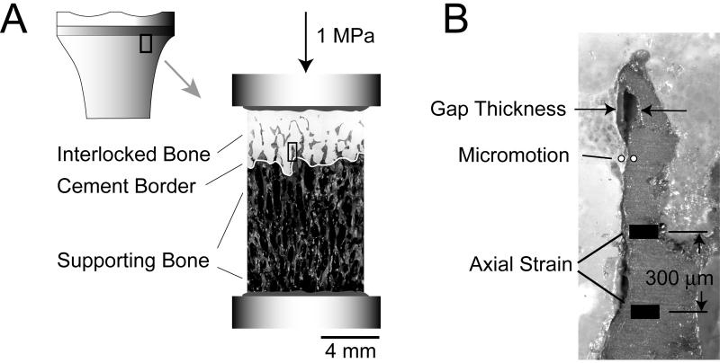
Figure 1.
Small segments of the cement-bone interface from below the tibial tray were sectioned (A) and loaded in axial compression to 1 MPa. The gap thickness, micromotion, and axial strain were measured at 50 randomly selected points along the trabeculae-cement interface (B). The gap thickness and micromotion measures were made across the trabeculae-cement interface. The axial bone strain was measured across a 300 μm gage length in the axial direction. All three measurements were made at coincident sampling points along the interface; they are shown here at three distinct locations for clarification purposes. The selected image in panel B is taken from the region of interest indicated by the black rectangle in the center image.

