
Fig. 8.
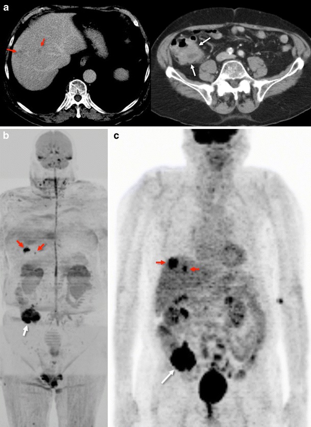
Whole-body diffusion imaging for tumour staging. Conventional portal-phase contrast-enhanced images (a) demonstrated two liver metastases (left – red arrows) and a tumour in the cecum (right - white arrow). Whole-body-diffusion-weighted image with inverted gray scale (b) depicted both focal liver lesions (red arrows) and the tumour in the cecum (white arrow) that showed restriction of diffusion. 18F-FDG-PET image (c) evidenced similar findings