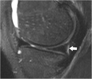
Fig. 15.

Coronal T2-weighted fat-suppressed fast spin echo image in a 21-year-old male patient shows the normal medial posterior femoral recess or medial gastrocnemius bursa with a small amount of fluid (arrow), which should not be diagnosed as a meniscocapsular separation
