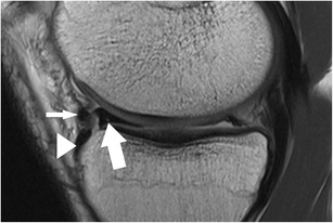
Fig. 21.

Sagittal proton-density–weighted fast spin echo image of the left knee in a 22-year-old male patient with a bucket handle tear of the medial meniscus shows the displaced anterior horn (arrow) which lies posterior to its root insertion (arrowhead). Note the relatively thick intermeniscal ligament (small arrow) that normally connects both anterior horns of the menisci as well as the small non-displaced remnant of the posterior horn
