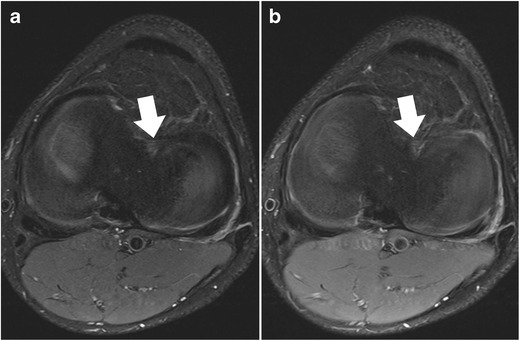
Fig. 3.

Set of two axial proton-density–weighted fat-suppressed fast spin echo images in a 42-year-old female patient, acquired at 2.5 mm slice thickness (a) and 5 mm slice thickness (b). With thinner slices, the meniscal root ligaments of the anterior horn of the medial meniscus (arrows) are much better appreciated. With thicker slices, these ligaments are barely visible, mainly due to partial volume effects
