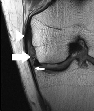
Fig. 6.

Coronal proton-density–weighted fast spin echo image of the right knee in a 25-year-old male patient with a traumatic osteochondral defect at the medial femoral condyle shows displacement of the medial meniscus and complete rupture of the meniscofemoral ligament (arrow). The more superficial medical collateral ligament (arrowhead) shows only mild abnormalities with thickening at its femoral insertion. Note the normal meniscotibial ligament (small arrow)
