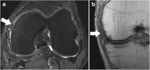
Fig. 8.

Axial proton-density–weighted fat-suppressed fast spin echo image (a) of the left knee in a 56 years old male patient shows an extra-articularly displaced fragment (arrow) of the medial meniscus. Coronal proton-density–weighted fast spin echo image (b) confirms the diagnosis and shows that the flapped portion (arrow) is still in continuity with the meniscus remnant (arrow)
