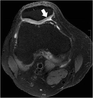
Fig. 9.

Axial proton-density–weighted fat-suppressed fast spin echo image of the right knee in a 40-year-old male patient acquired at 7 Tesla (Philips Healthcare, Best, the Netherlands) using a dedicated 28-channel transmit-receive knee coil shows a small articular cartilage fissure at the patella (arrow). Image in-plane resolution was 360 × 360 μm
