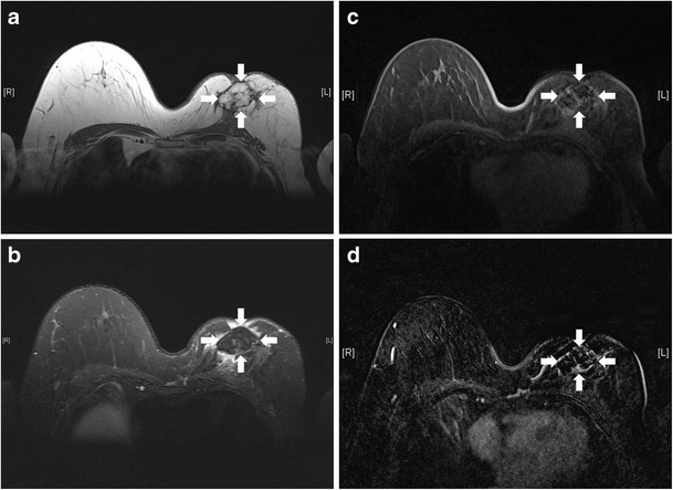
Fig. 25.

Fat necrosis after autologous fat implant. T1-weighted (a), T2-weighted, fat-saturated (b), and T1-weighted, fat-saturated (c) axial MR images show a multilobulated lesion (arrows) with signal identical to fat in the left breast. Mild perifocal edema is noted (b). T1-weighted, post-contrast axial MR image with subtraction (d) shows the non-enhancing nature of the fatty masses. A thin rim of enhancement surrounding them is likely due to post-operative changes
