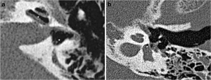
Fig. 10.

a Perilymphatic fistulae: CT axial views showing a perilymphatic fistula, highlighted by a pneumolabyrinth (white stars). Perilymphatic liquid has leaked into the middle ear. Massive pneumolabyrinth was seen 1 month after a translabyrinthine fracture, with air in the perilymphatic space (scala vestibuli and the vestibule) b Example of pneumolabyrinth without temporal bone fracture (white star)
