Fig. 13.

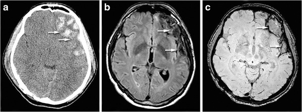
Brain haemorrhage in a patient with contralateral auditory agnosia. CT view (a) showing left fronto-temporal hemorrhagic injury. FLAIR sequence (b) and susceptibility-weighted imaging (c) showing the cortical damage in particular in the superior temporal gyrus and in the basifrontal region (white arrows)
