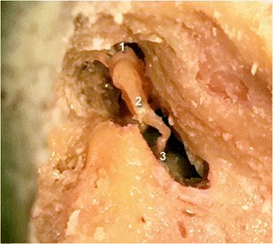
Fig. 3.

Cadaveric view showing a normal incudo-malleolar joint on the left side and a normal incudo-stapedial joint on the right side. As on the 3-D CT views, the malleus (1) and the incus (2) are in close contact. The incus is also in continuity with the stapes (3)
