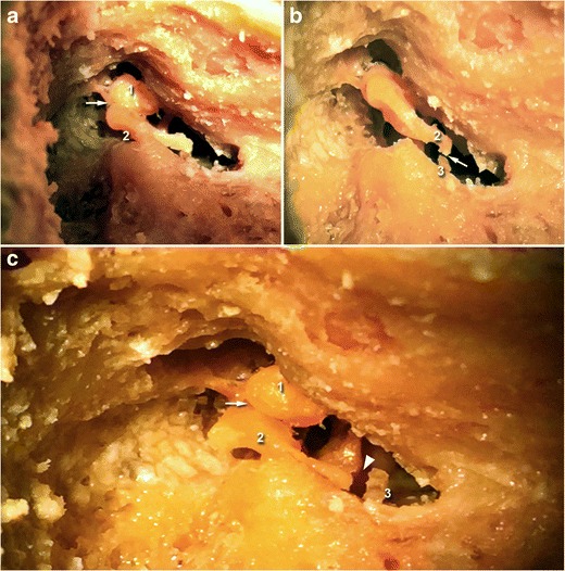
Fig. 6.

Cadaveric view showing incudo-malleolar luxation (white arrow, upper left) and incudo-stapedial luxation (white arrow, upper right). On the third image, both joints are dislocated (white arrow and white arrowhead). The malleus is annotated (1), the incus (2), and the stapes (3)
