. 2016 May 10;59(3):219–226. doi: 10.3340/jkns.2016.59.3.219

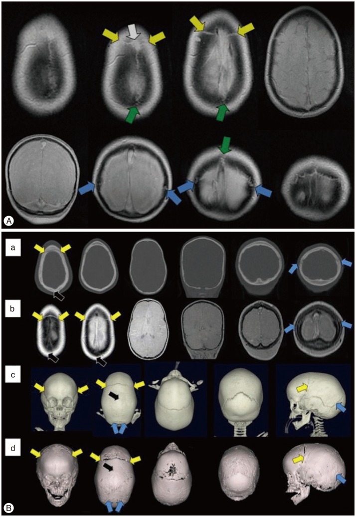
Fig. 3. Magnetic resonance imaging (MRI) findings of normal cranial sutures and craniosynostosis of children. A : Axial (top row) and coronal (bottom row) "Black Bone" imaging show normal patent cranial sutures as hyperintensity distinguished from the signal void of the cranial bones (Coronal suture : yellow arrow; Lambdoid suture : blue arrow; Sagittal suture : green arrow, visible anterior fontanelle : white arrow). B : Images of axial and coronal CT (a), axial and coronal "Black Bone" MRI (b), 3-D CT (c), and 3-D "Black Bone" MRI (d) show sagittal synostosis (Coronal suture : yellow arrow; Lambdoid suture : blue arrow; the site of synostosis : black arrow). Adapted from Eley KA, et al., Eur Radiol 24 : 2417-2426, 20146) with permission from Springer.

Fig. 3