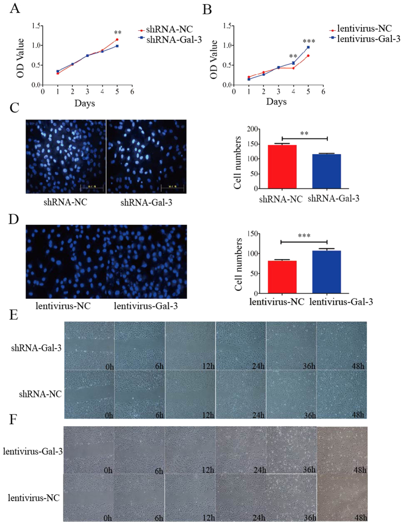
Figure 2. Cell proliferation and migration analyses of Gal-3-knockdown and -overexpressing BM-MSC lines in vitro.
Gal-3 enhanced the proliferation and migration of BM-MSCs. The proliferation rate of shRNA-Gal-3 cells was lower than that of shRNA-NC cells on day 5 (A), and the proliferation rate of lentivirus-Gal-3 cells was higher than that of lentivirus-NC cells on day 4 (B). Migrated Gal-3-knockdown (C) and -overexpressing (D) BM-MSCs in transwell chambers were stained with Hoechst 33342 and quantified (n = 3). Wound healing of Gal-3-knockdown (E) and -overexpressing (F) BM-MSCs was examined under a microscope. Knockdown of Gal-3 significantly inhibited the migration of BM-MSCs, and overexpression of Gal-3 significantly enhanced the migration of BM-MSCs in vitro. Magnifications: 20x (C,D) and 4x (E,F). Error bars represent the SEM (**p < 0.01, ***p < 0.0001).

