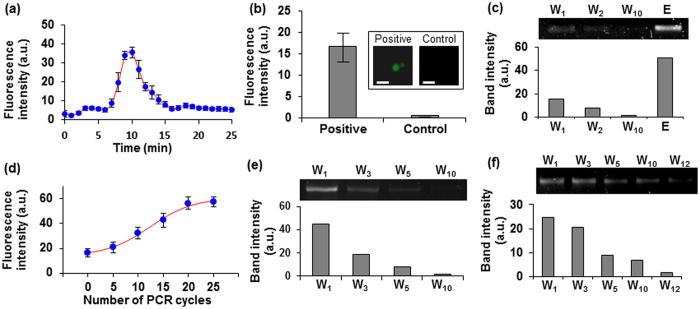
Figure 3. Characterization of electrophoretic transfer and bead-based PCR amplification of target-binding oligonucleotides.
(a) Fluorescence intensity measurements at the center of the gel-filled interconnection channel as fluorescently labeled IgE-binding oligonucleotides migrated from the selection chamber to the amplification chamber. (b) Fluorescence measurements of IgE-binding strands that were electrophoretically transferred to and captured by bead-immobilized reverse primers in the amplification chamber. Scale bars: 100 μm. (c) Gel electropherogram of oligonucleotides targeting the BA-glucose mixture eluted from the chip, following their electrophoretic transfer to, capture by, and subsequent thermal release from bead-immobilized reverse primers in the amplification chamber. (d) Fluorescence intensity of IgE-binding oligonucleotides amplified on beads following different numbers of PCR cycles. Affinity selection of PCR-amplified oligonucleotides against (e) IgE and (f) the BA-glucose mixture. Lane W: wash and Lane E: elution.

