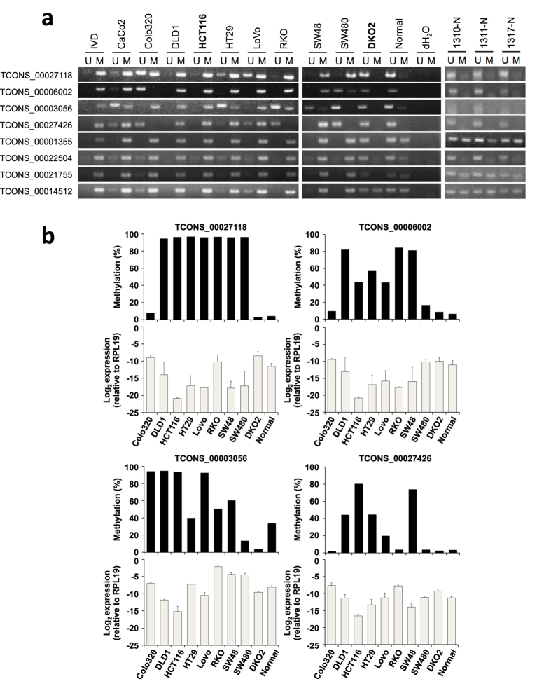
Figure 3. DNA methylation and expression of lncRNA genes in CRC cells.
(a) Methylation-specific PCR analysis of the promoter CGIs of the indicated lncRNA genes in CRC cell lines, a normal colonic tissue from a healthy individual (24 yo), and normal colonic tissues from CRC patients (1310-N, 74 yo; 1311-N, 75 yo; 1317-N, 79 yo). Bands in the “M” lanes are PCR products obtained with methylation-specific primers. Those in the “U” lanes are products obtained with unmethylation-specific primers. In vitro-methylated DNA (IVD) serves as a positive control. (b) The relationship between DNA methylation and expression of lncRNA genes in CRC cells and a normal colonic tissue. Shown are the results of bisulfite pyrosequencing (black bars) and quantitative RT-PCR (white bar) analysis of the four selected lncRNA genes. RT-PCR results were normalized to the internal RPL19 expression.

