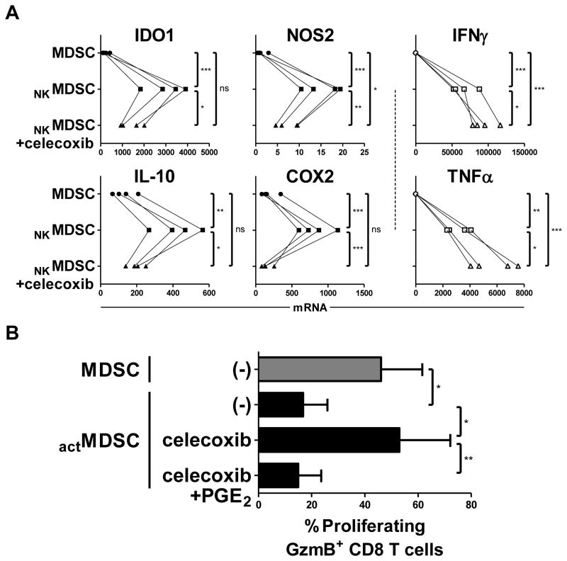
Figure 4.
Type-1 effector cell-driven hyperactivation of MDSCs requires the intact COX2-PGE2 axis. (A) Expression of IDO1, NOS2, IL10, COX2, IFNγ, and TNFα in OvCa ascites-isolated MDSCs cultured for 24 h with or without IL18/IFNα-activated NK cells (NKMDSC), in the additional presence or absence of celecoxib (COX2 inhibitor). Data are expressed as ratios between the expression of individual genes and HPRT1, and represent 4 independent patients. (B) Percentage of proliferating GzmB+ naïve CD8+ T cells following 4 d activation with anti-CD3/CD28 antibodies in the presence of resting or IFNγ/TNFα-activated OvCa ascites-isolated MDSCs (actMDSC), in the additional presence of celecoxib (COX2 inhibitor) and/or exogenous PGE2. Data represent the mean (± SD) of 4 independent patients. *** P < 0.001, ** P < 0.01, * P < 0.05, ns: P > 0.05 compared to indicated group.

