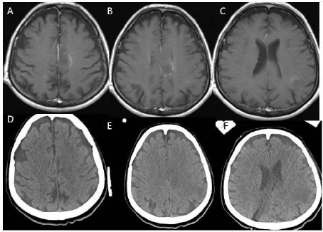
Figure 6.

(Group 2, case 19): A, B, C - Contrast-enhanced (Gadolinium) T1 weighted cranial MRI in axial sections showing weak hyperintense lesions on the left parasagittal region of the centrum semiovale and around left lateral ventricular area. D, E, F - Cranial CT without contrast in axial sections showing hypodense areas on the left parasagittal region of the centrum semiovale and around left lateral ventricular region corresponding with hypodense areas of cranial MRI.
